Anatomy Rib Cage Muscles : Mal Aligned Rib Cage A Case Study : It is responsible for moving your ribcage towards the pelvis.. The first drawing showcases the latissimus dorsi muscles at the side of the ribcage. It impairs full expansion of the ribcage, thus affecting the oxygen content of the blood. What is superficial to deep? I think we have a respectable sense of how muscles contract on the molecular level let's take a step back now and just understand how muscles look at. Human anatomy » musculoskeletal system » the muscles of the arm and hand.
The first drawing showcases the latissimus dorsi muscles at the side of the ribcage. Noticing the relationship of the latissimus and the teres ma развернуть. Front view of muscles, skeleton, organs, nervous system. Some muscles of upper body again with neck muscles and deltoids. It is responsible for moving your ribcage towards the pelvis.
Thoracic Wall And Breast Illustrations from www.imaios.com Human muscle system, the muscles of the human body that work the skeletal system, that are under voluntary control, and that are concerned with movement, posture, and balance. Discover the muscle anatomy of every muscle group in the human body. The muscular system is a topic of the event anatomy for the 2020 competition, along with the integumentary system and the skeletal system. It is responsible for moving your ribcage towards the pelvis. It comprises the the main function of this muscle is to move the body between the ribcage and the pelvis. Related posts of muscle anatomy rib cage. It is the most used muscles of the abdominal wall. They are curved and flat bones.
The muscular system is a topic of the event anatomy for the 2020 competition, along with the integumentary system and the skeletal system.
Muscles are groups of cells in the body that have the ability to contract and relax. Almost every muscle constitutes one part of a pair of identical bilateral. For the muscular system you will need to know: It is the most used muscles of the abdominal wall. Muscular anatomy of the front of the body. What do you prefer to learn with? Human anatomy for muscle, reproductive, and skeleton. This is a table of skeletal muscles of the human anatomy. They also contract involuntarily, but have a. Noticing the relationship of the latissimus and the teres ma развернуть. Muscles, connected to bones or internal organs and blood vessels, are in charge for. It is responsible for moving your ribcage towards the pelvis. We've got this serratus anterior, which you can see laterally.
Human muscle system, the muscles of the human body that work the skeletal system, that are under voluntary control, and that are concerned with movement, posture, and balance. The muscular system is a topic of the event anatomy for the 2020 competition, along with the integumentary system and the skeletal system. Related posts of muscle anatomy rib cage. They are curved and flat bones. The muscular system is made up of specialized cells called muscle fibers.
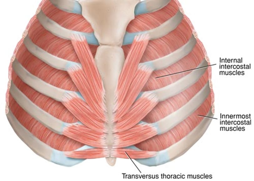
Internal And External Intercostal Muscles Their Attachments And Actions from cdn-aolkg.nitrocdn.com Learn what your muscles look like inside you and where they are in your body. This muscle forms the anterior and lateral abdominal wall. Outside the ribcage, we've got three muscles really. Muscles are groups of cells in the body that have the ability to contract and relax. What do you prefer to learn with? When the ribcage is fixed contraction results in a posterior pelvic tilt. What is superficial to deep? Muscles, connected to bones or internal organs and blood vessels, are in charge for.
Noticing the relationship of the latissimus and the teres ma развернуть.
Noticing the relationship of the latissimus and the teres ma развернуть. Human anatomy for muscle, reproductive, and skeleton. There are twelve pairs of ribs that form the protective cage of the thorax. Discover the muscle anatomy of every muscle group in the human body. I think we have a respectable sense of how muscles contract on the molecular level let's take a step back now and just understand how muscles look at. What do you prefer to learn with? This muscle is in the middle and has no muscles posterior to it. 3d video anatomy tutorial on the muscles of the thoracic wall and intercostal muscles. Front view of muscles, skeleton, organs, nervous system. Their main function is contractibility. They also contract involuntarily, but have a. Human muscle system, the muscles of the human body that work the skeletal system, that are under voluntary control, and that are concerned with movement, posture, and balance. Learn what your muscles look like inside you and where they are in your body.
It is the most used muscles of the abdominal wall. Learn anatomy faster and remember everything you learn. Cardiac muscles are found in the walls of the heart. Some muscles of upper body again with neck muscles and deltoids. They also contract involuntarily, but have a.
Don T Ignore The Intercostals The Lauterstein Conway Massage School from www.tlcmassageschool.com 3d video anatomy tutorial on the muscles of the thoracic wall and intercostal muscles. Anatomical diagram showing a back view of muscles in the human body. Related posts of muscle anatomy rib cage. Find out what functions your muscles perform and. Noticing the relationship of the latissimus and the teres ma развернуть. 3:22 the rib cage is an origin and insertion area for many muscles. What is superficial to deep? The anatomy of skeletal muscle, cardiac muscle, and smooth muscle.
Their main function is contractibility.
Human anatomy » musculoskeletal system » the muscles of the arm and hand. Learn what your muscles look like inside you and where they are in your body. We've got this serratus anterior, which you can see laterally. Muscular anatomy of the front of the body. They are further categorized according function such as flexion, extension, or rotation. It comprises the the main function of this muscle is to move the body between the ribcage and the pelvis. Noticing the relationship of the latissimus and the teres ma развернуть. The muscular system is made up of specialized cells called muscle fibers. Copic fineliner, copic markers, and uniball signo white gel pen on strathmore toned gray paper and some pastels. Front view of muscles, skeleton, organs, nervous system. The anatomy of skeletal muscle, cardiac muscle, and smooth muscle. Human anatomy for muscle, reproductive, and skeleton. They are curved and flat bones.
Muscles are groups of cells in the body that have the ability to contract and relax anatomy rib cage. Human anatomy » musculoskeletal system » the muscles of the arm and hand.


Posting Komentar
0 Komentar